Introduction: Management of the large gap in long bone fractures is a challenging problem after compound injuries. A novel technique called as Masquelet’s technique of “induced membrane formation”, is used to bridge a gap of more than 5 cm using bone cement as a spacer in first stage and autologous cancellous bone graft to fill the gap in second stage.
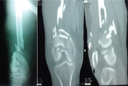
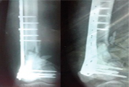
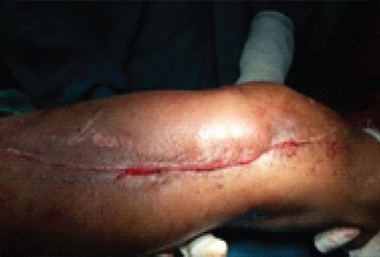
Case presentation: We present two different and difficult cases with bone defects after open injuries associated with long bone fractures in this paper. First case is a 50-year-old lady with grade IIIa open fracture right distal femur with intra-articular extension and bone loss. She underwent wound debriment, stabilization of the fracture with locking compression plate along with antibiotic cement spacer, which is removed latter and underwent bone grafting. Another is a 15-year-old boy with open grade IIIb fracture tibia and fibula (mid-distal third junction) of right leg, wound debridement and ankle spanning triangular external fixation was applied on same day and after two months, external fixation was removed due to florid infection and plaster of Paris was applied. Instead of the tedious and demanding treatment options like Ilizarov, a new technique described by Masquelet is used here. It uses bone cement as a spacer to fill the cavity to form pseudo-membrane around it and in the second stage autologous cancellous bone graft fills the gap of even more than 5 cm, to achieve union.
Conclusion: The membrane also secretes vascular and osteo-inductive factors to stimulate bone regeneration and also prevents resorption of the bone graft and achieves early fracture healing avoiding tedious options like bone transport in external fixator. By this two staged technique, union occurred clinically and radiologically in these two cases.


")